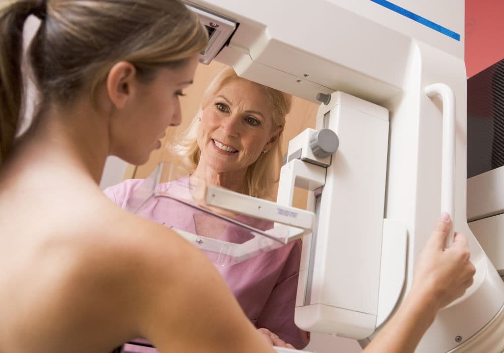

A mammogram is a specialized X-ray of the breast that can detect signs of breast cancer, such as lumps or abnormal tissue. It involves compressing the breast between two plates to obtain high-quality images. Mammograms are a crucial screening tool for women over the age of 40, as they can identify breast cancer in its earliest stages, often before symptoms are present.
Understanding Mammograms: What Are They?
Detecting Stage 1 Breast Cancer through Mammograms
Mammograms are an essential tool for detecting stage 1 breast cancer. This early stage of the disease is when the cancer is still localized and has not spread beyond the breast tissue. Mammograms are able to detect tiny lumps or growths in the breast that may not be noticeable through physical examination. They can also identify areas of calcifications or abnormal tissue that could be indicative of cancerous cells. By catching stage 1 breast cancer through mammograms, women have a much higher chance of successful treatment and survival. Regular mammograms for women over the age of 40, or those with a family history of breast cancer, are recommended as a proactive measure for early detection. While mammograms can be uncomfortable, the benefits of early detection and the potential to save lives make them an essential component of women's healthcare. Early detection through mammograms also means less invasive treatments and a higher likelihood of complete recovery.
How Mammography Can Aid in Early Breast Cancer Diagnosis
Mammography plays a vital role in the early detection of breast cancer, particularly Stage 1 breast cancer. Stage 1 breast cancer refers to cancer that is still localized within the breast and has not spread to nearby lymph nodes or other areas of the body. Mammograms can detect small, early-stage tumors that are not yet palpable during a physical examination. Early detection significantly improves prognosis and treatment options.
Recalled after a Mammogram: Common Reasons and What to Expect
Being recalled after a mammogram can be unsettling, but it is important to remain calm and understand that it is a precautionary measure. Common reasons for recall include unclear or inconclusive images, areas that require additional imaging for further evaluation, or the need for comparison with previous mammograms. If you are recalled, it does not necessarily mean you have breast cancer. Additional imaging or tests will help provide a more definitive diagnosis.
Distinguishing Cysts from Cancer on a Mammogram
When examining a mammogram, it can be challenging to distinguish between cysts and cancerous tumors. Cysts are fluid-filled sacs that are typically benign, whereas cancerous tumors are abnormal growths of cells that can spread and cause harm. One way to differentiate between the two on a mammogram is by examining the appearance of the mass. Cysts usually have a regular shape and smooth edges, while cancerous tumors often have irregular shapes and jagged edges. Additionally, the presence of calcifications within the mass can also indicate a higher likelihood of cancer. Another method for distinguishing between cysts and tumors is by conducting a fine-needle aspiration or a biopsy, which involves removing a sample of the tissue to examine it more closely. It's crucial for healthcare professionals to accurately identify the nature of the mass in order to provide the appropriate treatment and care for the patient, whether it be monitoring the cyst for changes or initiating necessary cancer treatment.
Interpreting Mammogram Findings and Identifying Abnormalities
When analyzing mammogram results, healthcare providers look for various abnormalities that may indicate breast cancer. One common finding is the presence of cysts, which are fluid-filled sacs and are usually benign. Cysts typically appear as round or oval shapes on a mammogram. Differentiating them from cancerous masses, which may appear as irregularly shaped clusters or spiky masses, requires careful evaluation by an experienced radiologist.
Deciphering Mammogram Results: What You Need to Know
Once your mammogram is complete, you will receive the results from your healthcare provider. Understanding these results is crucial for taking appropriate actions. The results may indicate normal findings, benign conditions, or abnormalities that require further evaluation. In some cases, additional tests such as ultrasound or biopsy may be recommended. It is essential to communicate with your healthcare provider to fully comprehend your results and any necessary follow-up steps.
In conclusion, mammograms are essential for the early detection and diagnosis of breast cancer. By understanding what mammograms are, their role in detecting Stage 1 breast cancer, reasons for recall, differentiating cysts from cancer, and interpreting mammogram results, you will be better equipped to navigate the screening process and follow-up evaluations. Regular mammograms can significantly improve breast health and increase the chances of successful treatment if breast cancer is detected.
Breast screening: guidance for image reading
Breast screening is an essential tool for detecting breast cancer early on, when chances of successful treatment are higher. Image reading is a critical aspect of breast screening, as it requires specialized knowledge and training to accurately interpret mammograms and other imaging tests. Guidance for image reading is crucial to ensure that healthcare professionals are equipped with the necessary skills and information to identify any abnormalities or potential signs of breast cancer. This guidance includes detailed protocols and standards for interpreting images, as well as ongoing education and support for image readers to stay updated on the latest advancements in breast screening technology. Additionally, ongoing quality assurance programs help to maintain the accuracy and consistency of image reading across different healthcare settings. Ultimately, providing comprehensive guidance for image reading in breast screening is essential for improving early detection and treatment outcomes for women at risk of breast cancer.
Imaging with breast implants
Imaging with breast implants can present challenges for healthcare providers. Traditional mammography may be less effective at detecting abnormalities in women with breast implants due to the implants obscuring the breast tissue. Therefore, specialized techniques such as displacement views and Eklund views may be used to improve the accuracy of the imaging. Additionally, women with breast implants may undergo alternative imaging methods such as MRI or ultrasound to screen for breast cancer or evaluate potential issues with the implants. It is important for healthcare providers to be knowledgeable about the specific considerations and techniques for imaging women with breast implants to ensure accurate diagnosis and appropriate treatment. Patients with breast implants should also inform their healthcare provider about the presence of implants to ensure that the most effective imaging methods are used for their particular situation. Overall, imaging with breast implants requires a tailored approach to accommodate the unique challenges and considerations associated with implant presence.
What is digital mammography?
Digital mammography is an advanced imaging technique used to screen for and diagnose breast cancer. It uses digital receptors and computers to create detailed images of the breast tissue, allowing for accurate detection of abnormalities such as tumors or masses. Digital mammography offers several advantages over traditional film mammography, including improved image quality, the ability to adjust and manipulate images for better visualization, and easier storage and transmission of images. This technology also allows for a quicker and more efficient screening process, leading to faster diagnosis and treatment. Digital mammography has become the gold standard for breast cancer screening and is recommended for all women, especially those with dense breast tissue or a high risk of developing breast cancer. It has revolutionized the way breast cancer is detected and treated, leading to earlier detection and improved outcomes for patients.
What Is Breast Cancer Screening?
Breast cancer screening involves the use of various tests and examinations to assess the presence of cancerous cells in the breast tissue. The goal of screening is to detect breast cancer at an early stage, when it is most treatable. The most common method of screening is through mammograms, which are X-ray screenings of the breast tissue. Other screening methods may include clinical breast exams by a healthcare provider and self-exams performed by the individual. Breast cancer screening is recommended for women starting at a certain age, typically around 40 to 50 years old, and should be done regularly as recommended by a healthcare professional. Early detection through regular screening can significantly improve the chances of successfully treating breast cancer and reducing mortality rates. It is important for individuals to discuss their risk factors and screening options with their healthcare provider to determine the best course of action for their specific situation.